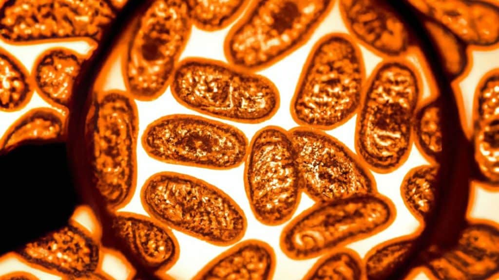
If you follow aging and longevity-related health news at all, you’re probably familiar with mitochondria. They’re essential to our energy metabolism and all sorts of different cell functions. This means it’s pretty important to know how well they’re working. A new imaging technique may allow us to do just that (https://longevity.technology/news/new-technique-for-studying-mitochondria-could-enhance-neurodegenerative-imaging/).
Every human has hundreds, if not thousands, of mitochondria in just one cell, and each mitochondria has its own distinctive structure. These structures can change depending on the specific function being performed but are also affected by outside stressors. Research has suggested Parkinson’s disease, Alzheimer’s disease and various cancers may all involve problems with the mitochondria.
Until now, viewing and analyzing the details of how mitochondria morph with different diseases has been difficult. We’re talking about very small and very complex structures where even subtle shifts can have a big impact. Now, an advanced microscopy technique known as cryo-electron tomography (cryo-ET) may make things just a little bit easier.
Cryo-ET doesn’t use light like other forms of imaging, relying on electrons instead. This allows for the creation of very detailed 3D models that can then be measured and analyzed in all sorts of ways. The scientists, based at the Department of Integrative Structural and Computational Biology at Scripps Research, used what they call a “surface morphometrics toolkit” to perform this analysis.
This toolkit was then used to assess every aspect of the mitochondria, from the inner and outer membranes to the gaps between them. First, it could be used to examine the mitochondria as they originally appeared, then it measured how the mitochondria responded to stress.
Endoplasmic reticulum stress is something that can happen to cells and is often associated with neurodegenerative diseases. When the scientists applied endoplasmic reticulum stress to the mitochondria and then analyzed the response with their new toolkit, they could see how the space between the membranes changed in size and how the inner membrane saw its usual curves reshaped.
Now the scientists hope to use their toolkit to examine even more types of mitochondrial stress. This will not only allow us to better understand the progression of various diseases; it could also help us plan treatments. If we want to test new drugs to try to “fix” the mitochondria, we may be able to see whether or not they work. It’s an important step forward.


